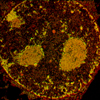
 Studying the dynamic properties of the genome and other nuclear
structures through time lapse microscopy of living cells can be used to
obtain information on the nature of structures or the execution of
important nuclear functions such as transcription, replication, and
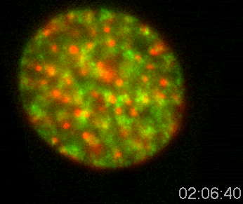
repair of DNA or processing messenger RNA. Most nuclear structures
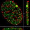
are not visible by traditional light microscopy. In order to study
structures or proteins within the living cell nucleus, it is necessary
to attach a fluorescent molecule that will cause the molecule to
fluorescence that can be imaged using fluorescence microscopy. In
most cases, this involves the construction of a synthetic version of a
nuclear protein that codes for a fusion of this protein with a protein
that emits fluorescence. The most commonly used fluorescent
protein tag is
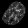
Green Fluorescent Protein (GFP). Two basic types of studies
can be performed. Object tracking experiments can be performed on
structures large enough to be detectable as single objects. For
example, an individual gene can be studied over time.
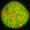
Molecular kinetic techniques, such as
fluorescence recovery after photobleaching (FRAP) and
fluorescence correlation spectroscopy (FCS) measure the diffusion of
molecules within the living nucleus. These studies can reveal the
relationship between visible structures and the individual molecules
that they contain
|