Source-Based Review Summary

The Nucleus as a Nexus of Proteostasis

A Critical Review of Protein Quality Control Mechanisms, Regulation, and Disease

8 major sections

Review

Explore the evidence and suggest corrections to this review.

Evidence-led review · 16 November 2026

Nuclear protein quality control: mechanisms and limits

Nuclear proteostasis includes folding assistance, organization of stressed proteins and their eventual refolding, export or degradation. These outcomes are distinct: a visible inclusion or the recruitment of a quality-control protein does not by itself establish that a substrate was refolded, destroyed or rendered harmless. The original review framed these processes as an interconnected network; the evidence below develops that framing with specific experimental systems and their limits.

Chaperones, disaggregation and proteasomal turnover

In budding yeast, den Brave and colleagues studied stress-induced nuclear protein inclusions and found that the nuclear J-domain protein Apj1 works with Hsp70 to solubilize substrates in a pathway coupled to their proteasomal turnover. This Apj1-dependent route was distinct from Hsp104-mediated disaggregation in their experiments. Loss of Apj1 uncoupled disaggregation from efficient clearance and was associated with accumulation of toxic soluble species. These results show why disappearance of a focus alone is an incomplete endpoint: one must determine what happened to its constituent proteins. The mechanism was established in yeast and must not be assumed to be identical in human nuclei. [1]

The ubiquitin–proteasome system can remove selected proteins, while molecular chaperones can support folding, solubility or delivery to degradation. A change in ubiquitin staining indicates altered localization or abundance of a label, not necessarily a measured change in substrate degradation rate. Proteasome inhibition can also create a strong proteotoxic stress of its own; conclusions should distinguish effects during inhibitor exposure from events after the inhibitor is removed. [1] [2]

Nucleolar stress and recovery

Frottin and colleagues observed stress-sensitive proteins entering the nucleolar granular component in mammalian cultured cells. In their system, recovery and extraction involved Hsp70, while prolonged stress impaired reversibility. The observations support a context-dependent protective role for nucleolar organization, not a conclusion that every nucleolus-associated inclusion is beneficial or that an imaging phenotype alone proves a particular material-state mechanism. [3]

Brunello and colleagues examined nucleolus-related inclusions after heat shock or proteasome inhibition, including experiments in H1299 cells. In the tested recovery conditions, inclusion clearance required UBA1 and nuclear proteasomes; HSP70, HUWE1 and the Pol I-dependent non-coding RNA IGS42 also contributed. UBA1 was required for recovery rather than for inclusion formation in that model. Autophagy/lysosome and CRM1-dependent export were not required for the particular clearance process tested. These findings should not be generalized to all inclusion types, stressors or cell states. [2]

Exchange with the cytoplasm and disease models

The nucleus is not an isolated protein-quality compartment. In autophagy-compromised human-cell models, Park and colleagues reported increased cytoplasm-to-nucleus movement of proteins and a contribution of nuclear proteasomes to their degradation. Their study also examined Huntington-disease cell models and reported altered nuclear transport in those models. These experiments support a compensatory connection between compartments under the tested conditions; they do not establish that the same route is protective in every disease, tissue or stage. [4]

How to interpret a quality-control experiment

Selected primary studies

  1. den Brave F et al. Chaperone-mediated disaggregation and clearance of intranuclear inclusions. Cell Reports 31:107680 (2020). PubMed 32492414. Evidence summarized from the abstract and selected figure descriptions.
  2. Brunello L et al. A nuclear protein quality control system for elimination of nucleolus-related inclusions. EMBO Journal 44:801–823 (2025; online 2024). PubMed 39690241. Evidence summarized from the abstract and selected figure descriptions.
  3. Frottin F et al. The nucleolus functions as a phase-separated protein quality control compartment. Science 365:342–347 (2019). PubMed 31296649.
  4. Park SJ et al. Nuclear proteasomes buffer cytoplasmic proteins during autophagy compromise. Nature Cell Biology 26:1691–1699 (2024). PubMed 39209961. Evidence summarized from the article abstract and selected reported results.
Overview

The maintenance of a functional proteome is a fundamental prerequisite for cellular life. This state of equilibrium, termed protein homeostasis or proteostasis, is governed by a complex, integrated network of pathways that regulate the synthesis, folding, conformational maintenance, trafficking, and degradation of all proteins within a cell. The proteostasis network (PN) ensures that proteins adopt and maintain their correct three-dimensional structures, are present at appropriate concentrations, and are localized to their correct subcellular compartments. However, the capacity of this network is finite and under constant challenge from genetic mutations, biosynthetic errors, and a host of environmental and physiological stressors. A failure to maintain proteostasis leads to the accumulation of misfolded, non-functional, and potentially toxic protein species, a pathological hallmark of numerous human diseases, including neurodegeneration, cancer, and the aging process itself.

Historically, research into protein quality control (PQC)—the surveillance arm of the proteostasis network—has been dominated by studies of the cytoplasm and the endoplasmic reticulum (ER). These compartments represent major sites of protein synthesis and folding, and our understanding of chaperone-assisted folding, the unfolded protein response (UPR), and ER-associated degradation (ERAD) has provided a foundational paradigm for the field. However, accumulating evidence has compelled a significant shift in focus toward the cell nucleus. Far from being a quiescent repository for the genome, the nucleus is now understood to be a highly active and critical compartment for PQC, equipped with a sophisticated and specialized toolkit to preserve its own proteome and, remarkably, to assist other compartments under stress.

Featured Figure
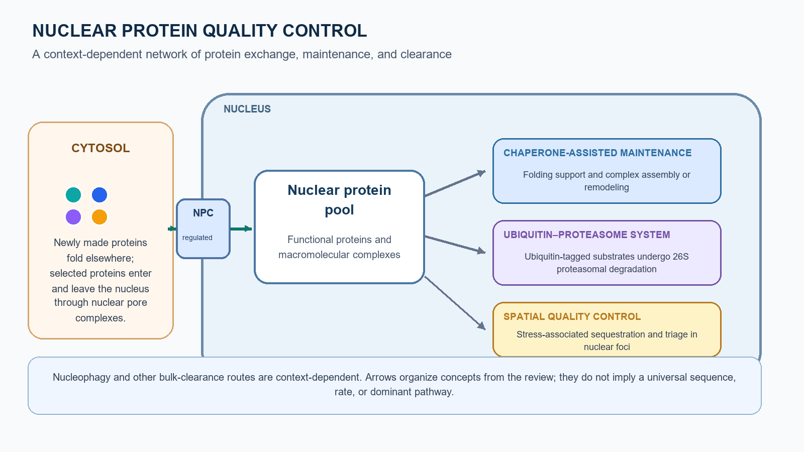
Diagram of nuclear protein quality control, showing protein exchange through nuclear pores, chaperone-assisted maintenance, ubiquitin-proteasome degradation, and stress-associated sequestration.

Figure 1. A context-dependent network of nuclear protein quality control.

Schematic summary of protein exchange, chaperone activity, proteasomal degradation, and spatial quality control discussed in this review. Nucleophagy and other bulk-clearance routes are context-dependent; arrows do not imply a universal sequence or dominant pathway. Not to scale.

Section 1

I. Introduction: The Imperative of Nuclear Proteostasis

The maintenance of a functional proteome is a fundamental prerequisite for cellular life. This state of equilibrium, termed protein homeostasis or proteostasis, is governed by a complex, integrated network of pathways that regulate the synthesis, folding, conformational maintenance, trafficking, and degradation of all proteins within a cell. The proteostasis network (PN) ensures that proteins adopt and maintain their correct three-dimensional structures, are present at appropriate concentrations, and are localized to their correct subcellular compartments. However, the capacity of this network is finite and under constant challenge from genetic mutations, biosynthetic errors, and a host of environmental and physiological stressors. A failure to maintain proteostasis leads to the accumulation of misfolded, non-functional, and potentially toxic protein species, a pathological hallmark of numerous human diseases, including neurodegeneration, cancer, and the aging process itself.

Historically, research into protein quality control (PQC)—the surveillance arm of the proteostasis network—has been dominated by studies of the cytoplasm and the endoplasmic reticulum (ER). These compartments represent major sites of protein synthesis and folding, and our understanding of chaperone-assisted folding, the unfolded protein response (UPR), and ER-associated degradation (ERAD) has provided a foundational paradigm for the field. However, accumulating evidence has compelled a significant shift in focus toward the cell nucleus. Far from being a quiescent repository for the genome, the nucleus is now understood to be a highly active and critical compartment for PQC, equipped with a sophisticated and specialized toolkit to preserve its own proteome and, remarkably, to assist other compartments under stress.

Section 2

II. The Unique Landscape of the Nuclear Proteome and its PQC Challenges

The necessity for a specialized and robust nuclear PQC network is dictated by the unique biophysical and functional characteristics of the nuclear environment, which present a distinct set of challenges compared to other cellular compartments.

The nucleoplasm is not a simple aqueous solution but a highly organized and exceptionally crowded environment. It is densely packed with macromolecules, including the entire genome organized into chromatin, a vast array of RNA molecules, and a high concentration of proteins. This molecular crowding creates a milieu where non-native proteins, which expose "sticky" hydrophobic patches, are at high risk of forming inappropriate and potentially toxic interactions with essential components like DNA, RNA, or functional protein complexes, thereby interfering with fundamental processes such as transcription, splicing, and DNA replication.

Key subtopics

  • The Crowded, Complex, and Charged Nuclear Milieu
  • The Challenge of Folded Cargo Import
  • A Comparative Analysis of Organellar PQC

Section 3

III. The Nuclear PQC Toolkit I: Molecular Chaperones and Protein Folding

Although the nucleus primarily imports proteins that have already been folded in the cytoplasm, it contains a versatile and essential suite of molecular chaperones. Their role extends far beyond the canonical function of refolding stress-denatured proteins. Within the nucleus, chaperones are critical for a host of constitutive processes, most notably the dynamic assembly, disassembly, and remodeling of the large macromolecular complexes that execute genomic functions. They act as crucial regulators of the "genomic interactome," ensuring its plasticity and responsiveness.

The Hsp70 family of chaperones, in conjunction with their Hsp40 (J-domain protein) co-chaperones, are ATP-dependent machines that bind to exposed hydrophobic regions on substrate proteins. In the nucleus, their functions are remarkably diverse. They play a key role in protecting labile structures, such as heterogeneous nuclear ribonucleoprotein (hnRNP) complexes, from stress-induced damage. A central regulatory role for Hsp70 is its involvement in the heat shock response (HSR). Under non-stress conditions, Hsp70 binds to and represses the master transcriptional regulator Heat Shock Factor 1 (HSF1). Upon proteotoxic stress, Hsp70 is titrated away to deal with the influx of misfolded proteins, liberating HSF1 to activate the transcription of protective genes, including more chaperones.

Key subtopics

  • The Hsp70 System (Hsc70/Hsp72)
  • The Hsp90 System
  • The Chaperonin CCT/TRiC

Section 4

IV. The Nuclear PQC Toolkit II: Degradation and Clearance Pathways

When molecular chaperones cannot rescue a misfolded or damaged protein, it must be eliminated to prevent its accumulation and potential toxicity. The nucleus is equipped with two major degradation systems that work in concert: the highly specific ubiquitin-proteasome system (UPS) for individual protein turnover, and the bulk-clearance pathway of nucleophagy for removing larger structures and aggregates.

The UPS is the principal mechanism for regulated protein degradation in eukaryotic cells and is highly active within the nuclear compartment. The system operates through a three-step enzymatic cascade that covalently attaches a chain of ubiquitin molecules to a substrate protein. The ubiquitin-activating enzyme (E1) activates ubiquitin in an ATP-dependent manner. The ubiquitin is then transferred to a ubiquitin-conjugating enzyme (E2). Finally, a ubiquitin ligase (E3) recognizes the specific substrate and catalyzes the transfer of ubiquitin to it. The formation of a polyubiquitin chain, typically linked through lysine 48 (K48) of ubiquitin, serves as a degradation signal (degron) that is recognized by the 26S proteasome, a large multi-catalytic protease complex that unfolds, deubiquitylates, and degrades the substrate into small peptides.

Key subtopics

  • The Nuclear Ubiquitin-Proteasome System (UPS): The Primary Degradation Engine
  • Nucleophagy: Bulk Clearance of Nuclear Debris

Section 5

V. Spatial Organization: Compartmentalizing Nuclear Proteotoxic Stress

A recurring theme in cellular PQC is the principle of spatial organization. Rather than allowing toxic, misfolded proteins to diffuse freely, cells sequester them into distinct, concentrated foci. This strategy offers several advantages: it mitigates the toxicity of aberrant species by isolating them from essential cellular machinery, it concentrates PQC factors like chaperones and ligases with their substrates to increase the efficiency of refolding or degradation, and it serves as a triage center to coordinate the fate of damaged proteins. In the nucleus, this spatial PQC is largely mediated by a variety of dynamic, membrane-less organelles or nuclear bodies, which are often formed through liquid-liquid phase separation (LLPS).

The formation of many nuclear bodies is now understood to be driven by LLPS, a physical process where multivalent interactions among proteins and/or RNA molecules lead to their spontaneous de-mixing from the surrounding nucleoplasm into a concentrated, liquid-like droplet. Proteins rich in IDRs or LC domains are particularly prone to undergo LLPS, and these domains are common among components of nuclear PQC bodies. The liquid-like nature of these condensates allows them to be highly dynamic, rapidly forming, dissolving, and exchanging components with the nucleoplasm in response to cellular needs and stress signals.

Key subtopics

  • Formation and Function of PQC Condensates
  • The Intranuclear Quality Control (INQ) Compartment
  • Nucleolus-Related Inclusions
  • Co-opting Pre-existing Nuclear Bodies for PQC

Section 6

VI. Regulation of Nuclear Proteostasis: From Cellular Stress to Systemic Coordination

The nuclear PQC network is not static; it is a highly dynamic system that must constantly adapt to fluctuating cellular conditions and organismal demands. This regulation occurs across multiple scales, from the rapid, local responses within the nucleus to cell-wide transcriptional programs and, as is now becoming clear, to systemic signals that coordinate proteostasis across the entire organism.

When a cell is confronted with proteotoxic stress, it activates intrinsic pathways to bolster its PQC capacity.

Key subtopics

  • Cell-Autonomous Stress Responses
  • The Emerging Frontier of Systemic Proteostasis

Section 7

VII. The Collapse of Nuclear Proteostasis: A Central Node in Aging and Disease

When the intricate network of nuclear PQC fails, the consequences are severe, leading to the accumulation of toxic proteins, disruption of essential nuclear functions, and cellular demise. This collapse of nuclear proteostasis is now recognized as a central pathogenic mechanism in a wide array of human disorders, from devastating neurodegenerative diseases and cancer to the fundamental process of aging itself.

A striking number of age-related neurodegenerative diseases are directly caused by the misfolding and aggregation of specific proteins within the nucleus. These are often referred to as proteinopathies, and the location of the toxic aggregates is key to their pathology.

Key subtopics

  • Neurodegeneration: A Story of Nuclear Protein Aggregation
  • Cancer: The Double-Edged Sword of Nuclear PQC
  • Aging and Premature Aging Syndromes

Section 8

VIII. Future Perspectives: Unresolved Questions and Therapeutic Opportunities

The study of nuclear proteostasis is a rapidly advancing field, propelled by technological innovation and a growing appreciation for its central role in health and disease. As our understanding deepens, new frontiers for investigation emerge, alongside promising avenues for therapeutic intervention.

Progress in cell biology is inextricably linked to the development of new tools to observe and manipulate cellular processes. The study of nuclear PQC is no exception.

Key subtopics

  • Methodological Frontiers: New Ways to See and Perturb Nuclear PQC
  • Key Unresolved Questions
  • Therapeutic Strategies: Targeting Nuclear PQC for Human Health